FTIR – new technologies, better questions
FTIR – new technologies, better questions
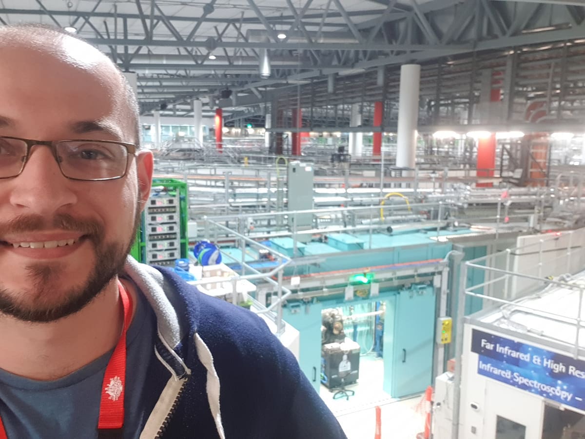
As scientists, we are always on the hunt for techniques and technologies that allow us to ask better questions about our research interests. I was fortunate enough to recently visit the Australian Synchrotron as part of my PhD programme. My supervisor Ross Graham’s (Curtin University) research group had received two grants of infrared radiation (IR) beamtime at the synchrotron; it was truly an incredible experience and privilege. When Ozgene provided me with an opportunity to pursue a PhD as a budding scientist on staff, I never expected to be in this position a mere 18 months later.

First, some background – what is a synchrotron?
Synchrotron as a light source
A synchrotron is a type of particle accelerator in which electrons are forced to travel in a near-circular pathway by magnets and emit electromagnetic radiation. This emission is known as synchrotron radiation; it is the result of bending the path of a charged particle. The Australian Synchrotron is a light source, which means that rather than accelerating particles in order to collide them and study their constituent parts, it is instead used to generate synchrotron radiation across the electromagnetic spectrum. This light is used for a variety of scientific and industrial applications – from protein crystallography and food analysis to medical imaging, and everything in between.
When it comes to my research field of biochemistry, the uses of the infrared spectrum of synchrotron radiation are of particular benefit, specifically Fourier transform infrared spectroscopy, or FTIR.
FTIR
FTIR is a spectroscopic method that uses the interferometric principle: the light beam is briefly split into two paths, one hits a mirror and the other hits the sample. These are then compared to determine the optical path difference. The FTIR instrument can be used to measure the absorption of light in a sample as a function of wavelength. The advantage of FTIR over other traditional spectroscopic techniques is its ability to collect data very rapidly, making it ideal for measuring samples that are difficult to prepare or that change quickly – such as live cells. FTIR can be used to identify functional groups in a molecule, study the conformations of molecules and investigate molecular interactions through the use of an FTIR spectrometer (or interferometer). FTIR is a very powerful tool for characterising organic and inorganic materials. FTIR spectroscopy works by shining infrared light through a sample and measuring the absorption of this light at different wavelengths. The resulting infrared spectrum can be used to identify the functional groups present in a molecule and to determine the structure of the molecule.
FTIR analysis
Analysis of the IR spectrum utilises the fact that different chemical groups in molecules absorb different wavenumbers (or have absorption bands) across the FTIR spectrum. This results in absorption peaks that can be used to relatively quantify the amounts of the chemical groups that are present.
Lifelines on the beamline
What I had not expected – yet was immensely grateful for – was the fantastic staff at the synchrotron. Mark and Pimm proved to be incredible founts of knowledge, with endless patience to explain everything: improving the signal-to-noise ratio, detailed explanations of the FTIR microscopes, sample preparation, IR radiation, optical path and different sampling technologies (such as attenuated total reflectance) as well as providing expert guidance on the use of an infrared spectrometer. They were instrumental to our success.
What is the better question?
I have been investigating the metabolism of a genetic knockout mouse that Ozgene generated and I found that there was a larger quantity of lipids in these animals’ liver sections when compared to wildtype. I observed this using a well-known histological stain: Nile Red, counterstained with DAPI. The liver is made of a repeating unit, called an acinus, that runs from a portal vein to a central vein – briefly, oxygen and nutrient-rich blood is supplied by the portal vein, which then diffuses across a field of hepatocytes towards the central vein. The distribution of lipids across the zones of an acinus is of biological relevance and I was able to quantify and spatially locate the lipids. However, I had no information about the chemical groups within the lipids. The ability to perform chemical identification on the lipids would further the understanding of where these lipids were coming from and how they were being used.
Here is where FTIR steps in: exploring serial sections from the same liver with IR spectroscopy yields the same spatial resolution, with the added depth of chemical groups. As the beam passes through, the sample absorbs light and generates an FTIR spectrum. Now I can analyse these chemical maps for their molecular bonds and gain an understanding of what makes up the lipid droplets observed in my Nile Red stains. This has been a goldmine of information and I can follow this within my samples to better understand the impacts of this gene knockout.
I strongly encourage scientists to explore newer technologies to expand their capabilities. It will allow them to ask better questions from the samples they have.
– Gorcin Kurejsepi, Organisational Capability Coach & PhD Candidate

